Super-resolution imaging has the capacity to distinguish and map structures that are smaller than the classical limit, typically a fraction of the wavelength. For ultrasound imaging, this means exploring features, such as blood vessels, in the micrometric range deep inside tissue. At the end of this course, students should be able to understand and reproduce super-resolution ultrasound imaging experiments, from data acquisition to image reconstruction, and apply such knowledge in their specific fields.
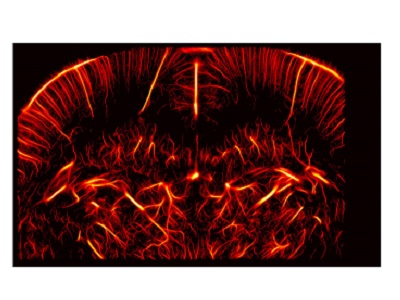
We first explore the fundamental aspects of imaging resolution in ultrasound. Various approaches to bypass the diffraction-limit with microbubbles and other agents are presented. More particularly, we discuss ultrasound localization microscopy, which has recently improved the resolution for vascular imaging by more than 10-fold. We present its various steps, including separation, localization and tracking, and compare different approaches. Specific elements such as temporal resolution, motion correction or volumetric imaging are considered. We then detail the applications of super-resolution ultrasound for brain, tumor, kidney, liver lymph nodes and peripheral vessels imaging, along with future perspectives in the clinical and preclinical context. The last part of the course will include hands-on image processing of in-silico and in-vivo open data with provided ultrasound localization microscopy algorithms.

